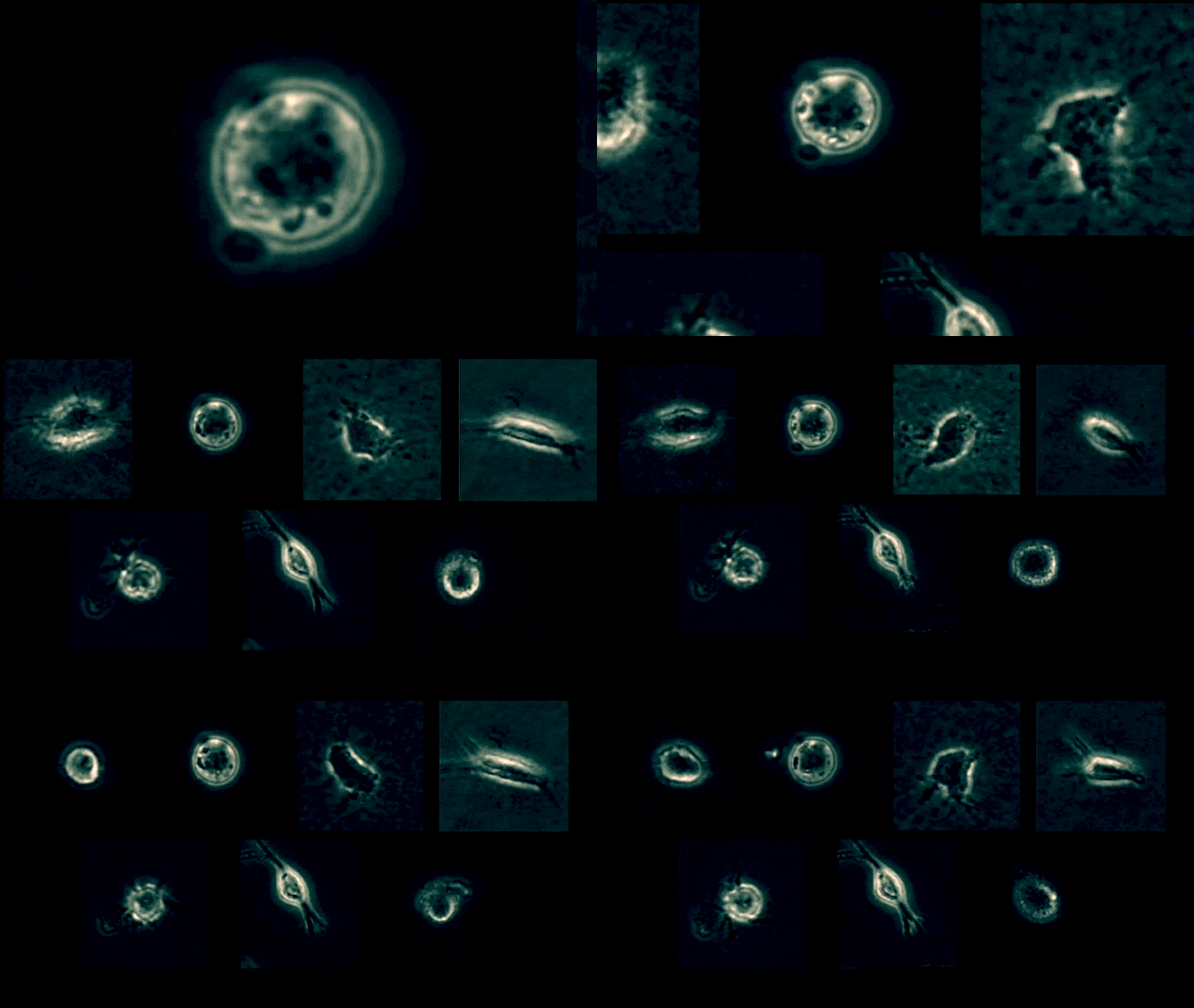
Scientists have developed a method to determine which tumor cells are most likely to metastasize efficiently to distant sites in the body. Assaf Zaritsky, now at Ben-Gurion University in Israel, and his colleagues in Gaudenz Danuser’s lab at UT Southwestern Medical Center designed a deep learning program that analyzes data from live phase-contrast imaging of melanoma cells taken from xenografts—mice implanted with patients’ tumor material. The program determined “the most representative morphological and behavioral properties of each melanoma cell and then demonstrated that this representation of the cell state can be used to predict in stage III melanoma excised from the lymphatic system the chances of progression to stage IV,” Zaritsky writes to The Scientist in an email. The scientists presented their “quantitative live cell histology” results at the American Society for Cell Biology...
Clarification (November 12): The article was updated to include the name of the lab where the work was performed.
Interested in reading more?


