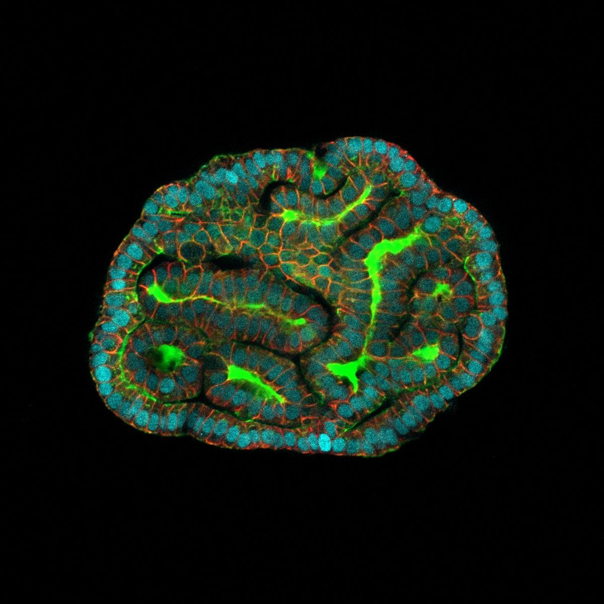
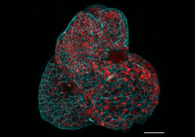
ABOVE: Researchers generated various epithelial organoids, including lungs, using progenitor cells found in amniotic fluid. Giuseppe Calà, Paolo De Coppi, Mattia Gerli
Moments after birth, a baby takes a first breath as the placenta, which has served as the fetus’ lungs during gestation, transfers responsibility to the baby's own organs. However, for patients born with congenital diaphragmatic hernia (CDH), a rare condition where the diaphragm fails to close, causing impaired lung development, entry into the world is more precarious.1 More severe cases of the disease lead to multiorgan damage, and approximately thirty percent of infants diagnosed with CDH never leave the hospital.2
Diagnostic imaging and genetic screens help clinicians catch congenital fetal diseases in utero, but models for studying organ development and disease progression are limited. Over the last decade, organoids have become an increasingly popular platform for modeling organ function and disease.3 However, the generation of fetal organoids is complicated by ethical and legal restrictions on the harvesting of the human tissues needed to generate the mini-organs.
Now, reporting in Nature Medicine, researchers generated fetal organoids using cells derived from human amniotic and tracheal fluids.4 These mini-organs offer a minimally invasive approach for disease modeling during an active pregnancy and may eventually inform the development of personalized prenatal interventions.
“Having access to the fetal tissue offers the possibility to model the fetal tissue while the baby is still in the womb,” said Mattia Gerli, a stem cell biologist at University College London (UCL) and study coauthor.
Scientists use patient cells to generate organoids that possess certain features and functions of the modeled organ while retaining the individual's genetic fingerprint. However, many of these platforms require lengthy dedifferentiation protocols to revert somatic cells into a state of pluripotency and then reprogram them to develop as another cell type.5 In contrast to organoids generated from pluripotent stem cells, primary organoids use tissue-specific stem cells or progenitor cells and therefore require minimal manipulation.3 While the organoid field is relatively advanced in terms of using adult tissues, researchers can only generate primary fetal organoids using tissue from terminated pregnancies. “This made it basically impossible to [generate organoids] compatible with the continuation of pregnancy, and therefore in a personalized medicine fashion,” said Gerli. To search for alternative sources of organoid building blocks, Gerli and his colleagues turned to amniotic fluid.
During gestation, the fetus floats in a protective pool of amniotic fluid.6 The yellowish liquid contains a concoction of nutrients and antibodies produced by the parent as well as less glamorous contributions from the fetus, including urine. It also includes fetal cells sloughed off during development, which doctors can extract and analyze for signs of disease.
“Those cells historically have been thought to be dead cells or cells that were shed from the lining of the amniotic fluid cavity,” said Shaun Kunisaki, a pediatric surgeon at Johns Hopkins University who was not involved in the study. Most amniotic fluid cells are epithelial, but scientists knew very little about these cell populations.3 “Everything changed when [Gerli] started to look at the single cell level at what happened in the amniotic fluid,” said Paolo De Coppi, a stem cell biologist and pediatric surgeon at UCL and study coauthor.

Gerli and his colleagues used single-cell RNA sequencing to characterize the amniotic fluid of 12 patients and discovered subpopulations of epithelial cells that expressed markers typical of progenitors for the lung, kidney, and small intestine. The researchers cultured the tissue-specific progenitor cells, fed them a chemical cocktail to support growth, and watched as they proliferated, differentiated, and self-organized into 3D epithelial organoids. The mini-organs shared some transcriptomic and protein features found in their tissues of origin. For example, lung epithelial cells that developed and differentiated in culture had elevated expression of airway markers compared to their nondifferentiated counterparts. Similarly, kidney epithelial organoids expressed markers associated with renal tubules, which are integral components of the kidneys’ filtration system.
Although the amniotic fluid contained cells from other tissues, the researchers could not grow them into organoids, suggesting that they lack progenitor capabilities. Other research groups have successfully grown fetal organoids from somatic cells floating around the amniotic fluid, and the mini-organs generated using this approach are more complex than the models developed by De Coppi and his team.7 However, reprogramming methods take up to 20 weeks to generate organoids. If the goal is to use organoids to inform prenatal interventions, timing is critical.
For the most serious cases of CDH, doctors perform prenatal surgery to implant a balloon that artificially blocks the trachea. This prevents lung fluid from escaping, leading to a buildup in pressure that helps promote lung development. De Coppi noted that the surgery is performed at around 25 weeks of gestation. However, doctors can only safely remove amniotic fluid starting around week 15, giving scientists a 10-week window to generate patient-specific organoids that scientists could use to test drugs that could further support lung growth when used alongside surgical interventions. Because the researchers use progenitor cells that do not require any reprogramming, they can generate the patient organoids in four to six weeks. “We lose complexity, but we gain time,” said Gerli.
To explore the usefulness of their platform in disease modeling, De Coppi and his team generated lung organoids using amniotic fluid from fetuses with CDH. The CDH mini lungs recapitulated some features of the disease, such elevated angiotensin 2 receptor (AT2) gene expression. Because they collected amniotic fluid both before and after surgical intervention, the researchers could compare organoids generated from the two time points. De Coppi and his team observed a reduction in the expression of the epithelial progenitor marker SRY-box transcription factor 9 (SOX9) following the procedure, indicating tissue maturation. However, due to a limited sample size, they did not perform within subject analyses.
Although sloughed off cells captured in the amniotic fluid offer a minimally invasive method for acquiring fetal tissues, Kunisaki said, “Whether or not [the cells] are a true reflection of what's going on within fetal development has always been a little bit controversial.” Kunisaki also noted that the study is limited to three cell types, but it’s often the interaction between various cell populations that is important for modeling organ development. “It's a little bit limited in terms of that, but regardless, the findings that they present are really promising,” said Kunisaki.
“The other possibility that [these organoids] open up is to study the development of the lung at a stage where the access to that tissue is very difficult,” said De Coppi. Given the varied legal restrictions on accessing fetal tissues, particularly beyond 22 weeks of gestation, Gerli said that their platform could provide scientists with a minimally invasive way to monitor the development of these tissues in later stages of pregnancy. “That is quite an important advance that this technology is bringing,” said Gerli.
The organoids still require clinical validation, but Gerli and De Coppi hope that their model will benefit patients down the road. “The aim with this is to actually be able to use the organoids as a platform for disease modeling and drug testing for these patients,” said Gerli.
A few drugs are commercially available, but patients respond differently to the treatments, which are currently only administered postnatally. Prenatal administration of these therapies could encourage proper diaphragm formation earlier in development. “That is one really intriguing possibility based on this kind of technology,” said Kunisaki.
- Zani A, et al. Congenital diaphragmatic hernia. Nat Rev Dis Primers. 2022;8(1):37.
- Politis MD, et al. Prevalence and mortality in children with congenital diaphragmatic hernia: A multicountry study. Ann Epidemiol. 2021;56:61-69.e3.
- Calà G, et al. Primary human organoids models: Current progress and key milestones. Front Bioeng Biotechnol. 2023;11:1058970.
- Gerli MFM, et al. Single-cell guided prenatal derivation of primary fetal epithelial organoids from human amniotic and tracheal fluids. Nature Medicine. 2024;30(3):875-887.
- Kim J, et al. Human organoids: Model systems for human biology. Nat Rev Mol Cell Biol. 2020;21(10):571-584.
- Underwood MA, et al. Amniotic fluid: Not just fetal urine anymore. J Perinatol. 2005;25(5):341-348.
- Kunisaki SM, et al. Human induced pluripotent stem cell-derived lung organoids in an ex vivo model of the congenital diaphragmatic hernia fetal lung. Stem Cells Transl Med. 2021;10(1):98-114.


