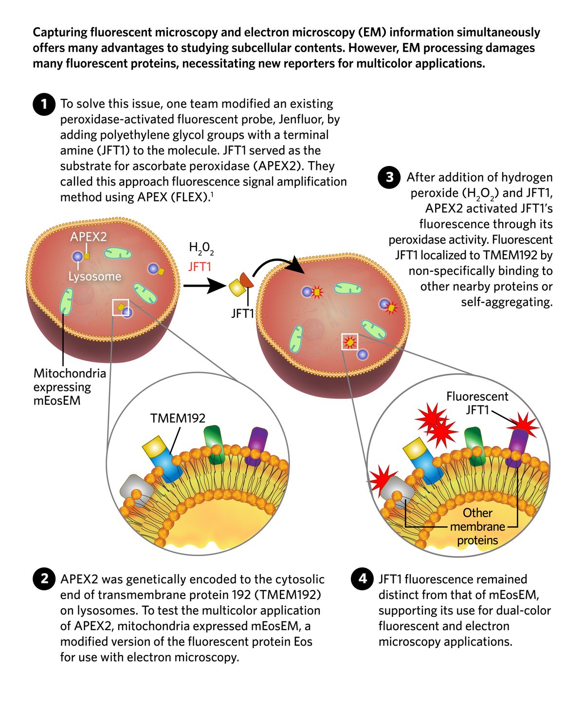
The first panel in a schematic of a modified probe, JFT1, used with a peroxidase (APEX2). APEX2, a yellow square, is shown to be on lysosomes, while Mitochondria are shown green with mEosEm, a fluorescent protein. The second panel is an insert of the lysosomal membrane with APEX2 bound to a blue lysosomal transmembrane protein. A grey, a green, and a purple membrane protein are also present. In the third panel, JFT1 has reacted with hydrogen peroxide and APEX2 to create red fluorescence near APEX2 that remains separate from the green mitochondria. The final panel is an insert showing the lysosomal membrane with activated JFT1, depicted as red spiky shapes, bound to the three other nearby membrane proteins or itself.
© istock.com, lvcandy, KKT Madhusanka; Designed by Erin Lemieux