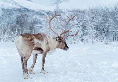
ABOVE: Reindeer shed and regrow their antlers without scars. Understanding this process can provide important insights to advance the field of regenerative medicine. ©iStock, RelaxFoto.de
With their hooves crunching over fallen leaves, reindeers dash into white, snowy landscapes—and folklore—as Christmas approaches. These majestic creatures, with their thorny headgear, play a bigger role than merely serving as Santa’s personal chauffeurs in legends; they are also important models to study a biological superpower.
Reindeer, along with their cousins from the deer family Cervidae, can shed and regenerate an entire organ. Both male and female reindeer are capable of regrowing antlers, unlike other deer species where this phenomenon is unique to male animals. Deer lose their antlers and regrow them at an unprecedented speed over the next few weeks.1,2 During this phase, specialized skin called velvet covers the antlers, which is shed a few weeks later, leaving behind bony antlers. The animals drop these structures again a few months later, beginning a new round of antler regeneration. Despite the repeated shedding and regeneration, deer regrow their antler skin flawlessly during each cycle without leaving behind any scars.

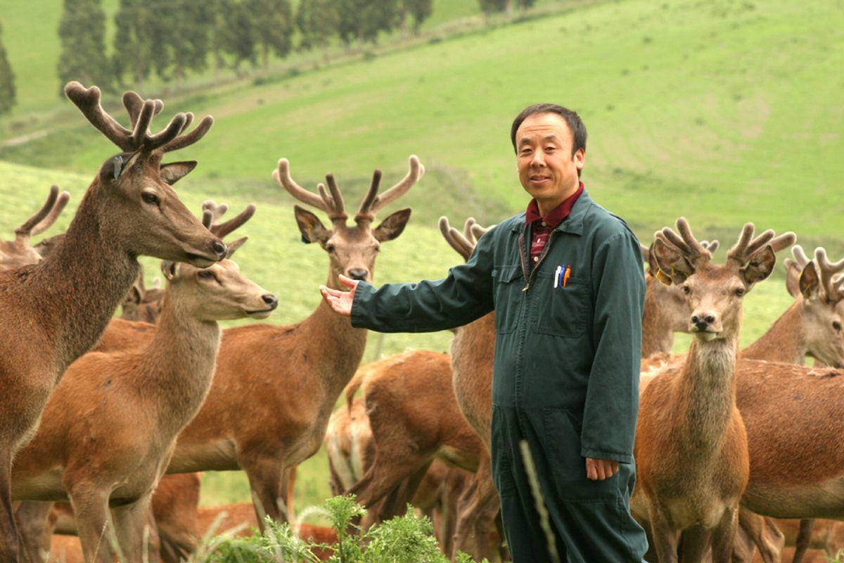
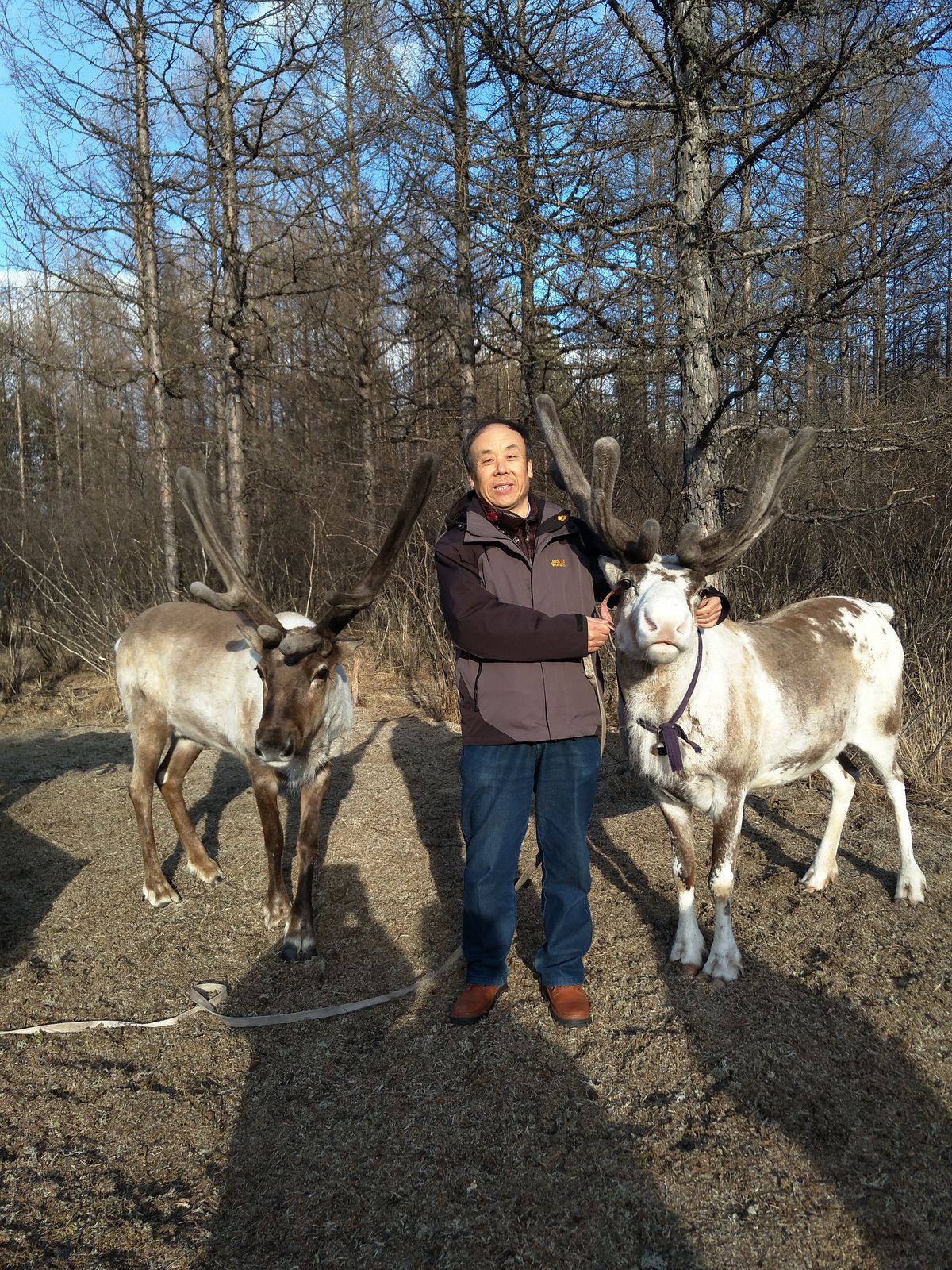
When Chunyi Li, an antler biologist at Changchun Sci-Tech University, started studying deer 40 years ago, he was struck by their ability to drop and regenerate antlers and immediately realized the applications of studying this process. “I started to think I should work in the field. And in the future, I may be able to help people to grow back their amputated limb or legs or arms,” said Li.
Although regenerating amputated limbs is still a distant dream, Li and researchers have obtained important insights about organ regeneration from studying the cellular and molecular processes behind tissue renewal and repair in deer. These insights can lay the foundation for better wound and tissue repair therapies, paving the way for advancing the field of regenerative medicine.
Deer As a Model to Study Regeneration
Members of the deer family are not the only mammals with regenerative abilities. Skin injuries in spiny mice heal without leaving any scars.3 Holes punched into the ear of spiny mice fully close with newly formed blood vessels, cartilage, muscle, and nerve fibers. But these rodents cannot regenerate an entire organ, said Li. “Antler is the only mammalian organ that once lost can fully grow back.”
This is even more remarkable given that antlers are complex organs, said Jeff Biernaskie, a regenerative biologist at the University of Calgary. “The organ consists of nerves, massive blood supply, bone, and cartilage.”
Deer are also a valuable platform to study wound healing. “Deer are similar to humans in that their skin is sort of tethered to underlying fascia muscle. They are also really fascinating because they are the only large animals that are able to [regenerate tissue].”
In addition to this, reindeer exhibit both regenerative and non-regenerative types of healing; antler velvets heal without forming a scar, while wounds in their back skin create scars.4 Reindeers mimic human scars more closely compared to animals like mice and rats which are commonly used to study scarring. “And so not only is it a great model of regeneration, but it's also a pretty good model of scar formation,” said Biernaskie.
Scientists have hypothesized that studying these unique properties of the deer family could point the way towards the biological mechanisms that can activate regenerative healing in other animals. To exploit the biology behind regeneration for medical applications, researchers must first understand which cells were involved in the process.
Stem Cells Aid Antler Regeneration
During the early 2000s, Li and his team focused on identifying the different types of cells in antlers. They used microscopy methods to pinpoint a population of cells present in antlers during regeneration.5,6
When the team deleted these cells in some deer, the animals showed no signs of antler regeneration throughout the season. The team transplanted this cell population elsewhere in the body of some deer and observed antler growth there, indicating that these cells were crucial for antler regeneration.7
Li and team then focused on characterizing this cell population. Using several cellular and molecular techniques, the team identified these cells as antler stem cells (ASCs).8 Li’s team recently carried out RNA sequencing of regenerating antlers to assess the origins of these ASCs.1 They identified a population of progenitor cells that gives rise to ASCs; these progenitor cells can differentiate into both bone and cartilage cells, which are important components of antlers.

To understand the mechanism behind ASC-induced regeneration, researchers studied the factors secreted by these cells. They observed that exosomes—membrane-enclosed structures—derived from ASCs prolonged the proliferation of human bone marrow stem cells in culture.9 These findings indicate that exosomes derived from these cells could be used to develop therapeutics that encourage regenerative healing.
Biernaskie believes that deer are valuable for studying more than just entire organ regeneration. He and his team investigate reindeers’ ability to exhibit both skin regeneration and scarring to understand the biological differences underlying the two types of healing.4 Using RNA sequencing and proteomics, the team found that wounds in velvet skin, which show scarless healing, lead to the activation of different immune cells compared to wounds in back skin, which form scars. Wounds on mice skin, which normally leave scars, healed without scarring when the researchers mimicked signaling pathways active in antler velvet wounds. These results highlight that insights obtained from the deer model could have potential applications in regenerative healing in other mammals.
Next, the team will employ a pig model to study which of the candidate pathways identified from reindeer could hold true across species. “Because if they work in mice, they work in pigs, then [it is] very likely that they're also going to work in humans,” said Biernaskie.
Future of Deer Models: Hit or Miss?
Although they can provide important clues into mammalian regenerative processes, deer are not widely studied. Both Li and Biernaskie attributed this to logistical difficulties. Deer handling requires experts in large animal veterinary care and wildlife medicine. Studying deer also requires facilities that are built to handle such large animals. This makes it very expensive, said Li.
There are also biological limitations to the model, which is not suitable for transgenic experiments, which are easier to perform in mice or other common lab animals, said Biernaskie. To circumvent this complication, researchers must first identify candidate regeneration-related genes in reindeer, followed by manipulating these genes in mouse models to validate their effects.
Moreover, velvet skin covers antlers for only about three months in a year, which gives researchers limited time to work on it. As a result, Biernaskie and team spent a significant time studying differences between the velvet and back skin wound healing in reindeer. “We worked on that particular project for almost 10 years before we published it,” said Biernaskie.
Despite these obstacles, the researchers believe that the deer is a useful model that can offer a lot of insights. “We're very open to collaborating with other groups that may not have access to, but have questions that they like to test in that system,” said Bierkaskie. “So, we can start to really exploit this [model] to its fullest potential.”
However, he does not believe that deer will enter mainstream animal models among researchers worldwide anytime soon, mainly due to the logistical challenges of working with such large animals. In such cases, studying other regenerative animals—including planaria and axolotls—can be helpful. This in combination with studying deer can provide important information about different strategies to enable tissue regeneration in the animal kingdom. “We're going to really start piecing this together over the next decade, and new therapies will start to arise.”
- Qin T, et al. A population of stem cells with strong regenerative potential discovered in deer antlers. Science. 2023;379(6634):840-847.
- Landete-Castillejos T, et al. Antlers—Evolution, development, structure, composition, and biomechanics of an outstanding type of bone. Bone. 2019;128:115046.
- Gaire J, et al. Spiny mouse (Acomys): An emerging research organism for regenerative medicine with applications beyond the skin. NPJ Regen Med. 2021;6(1):1-6.
- Sinha S, et al. Fibroblast inflammatory priming determines regenerative versus fibrotic skin repair in reindeer. Cell. 2022;185(25):4717-4736.e25.
- Li C, et al. Morphological observation of antler regeneration in red deer (Cervus elaphus). J Morphol. 2004;262(3):731-740.
- Li C, et al. Histological examination of antler regeneration in red deer (Cervus elaphus). Anat Rec A Discov Mol Cell Evol Biol. 2005;282A(2):163-174.
- Li C, et al. Identification of key tissue type for antler regeneration through pedicle periosteum deletion. Cell Tissue Res. 2006;328(1):65-75.
- Li C, et al. Adult stem cells and mammalian epimorphic regeneration-insights from studying annual renewal of deer antlers. Curr Stem Cell Res Ther. 2009;4(3):237-251.
- Lei J, et al. Exosomes from antler stem cells alleviate mesenchymal stem cell senescence and osteoarthritis. Protein Cell. 2022;13(3):220-226.


