Developmental Biology

A Matter of Molecular Attraction
Mariella Bodemeier Loayza Careaga, PhD | Sep 16, 2024 | 2 min read
While studying the metabolism of the developing chick embryo, Marià Alemany Lamana’s team acted quickly to avert an error.

Stem Cell-Based Embryo Models Add a Dimension to Developmental Biology
Shelby Bradford, PhD | Sep 13, 2024 | 10+ min read
Studying human embryonic development is complicated for several reasons. Models derived from pluripotent stem cells representing distinct stages offer a path to studying this process.
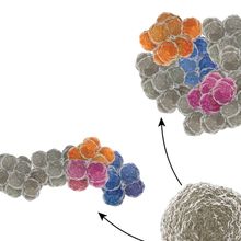
Infographic: The Many Paths to Stem Cell-Based Embryo Models
Shelby Bradford, PhD | Sep 13, 2024 | 5 min read
Culturing pluripotent stem cells to replicate different stages of embryonic development allows researchers to more easily investigate this period.

An Ode to Stem Cells
Meenakshi Prabhune, PhD | Sep 13, 2024 | 3 min read
Leveraging the versatility of stem cells allows researchers to advance science across multiple disciplines.
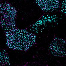
Introducing a New Version of the Cell Cycle
Maggie Chen | Sep 6, 2024 | 4 min read
Scientists have identified a new variant of the cell cycle that could provide insight into how diseases like cancer occur.
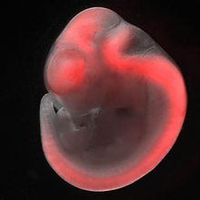
Dynamic Enhancers Orchestrate Development
Mariella Bodemeier Loayza Careaga, PhD | Sep 2, 2024 | 2 min read
Evgeny Kvon leverages transgenic models and genomic techniques to uncover the ways enhancers control the transcription of genes.
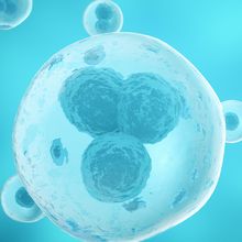
Study Reveals a Cell-Eat-Cell World
Aparna Nathan, PhD | Aug 13, 2024 | 3 min read
From normal vertebrate development to tumor cell cannibalism, cell-in-cell events occur in many different contexts across the tree of life.
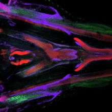
Unveiling the Secrets of Head and Face Formation
Mariella Bodemeier Loayza Careaga, PhD | Aug 1, 2024 | 5 min read
Samantha Brugmann illuminates the cellular and molecular factors that contribute to the formation of craniofacial structures.
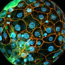
Unraveling the Complex Mysteries of Embryonic Beginnings
Shelby Bradford, PhD | Jul 4, 2024 | 6 min read
Magdalena Zernicka-Goetz followed the aesthetic allure of the embryo to better understand the start of development.

A Protein-Sensing Molecular Switch Alters Facial Features
Shelby Bradford, PhD | Jun 24, 2024 | 3 min read
The mTORC1 signaling pathway senses nutritional information and influences craniofacial development in mice.
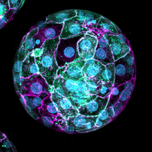
The First Two Cells in a Human Embryo Contribute Disproportionately to Fetal Development
Shelby Bradford, PhD | May 13, 2024 | 4 min read
A research team showed that, contrary to current models, one early embryonic cell dominates lineages that will become the fetus.
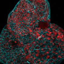
Fetal Organoids Generated From Human Amniotic Fluid
Danielle Gerhard, PhD | May 3, 2024 | 6 min read
A minimally invasive strategy for creating fetal organoids could facilitate precision medicine in the womb.
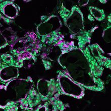
Growing Milk-Secreting Mammary Organoids
Sneha Khedkar | Mar 29, 2024 | 3 min read
Mammary organoids derived from mouse embryonic stem cells could offer clues into mammary gland developmental origins and help researchers study breast cancer.
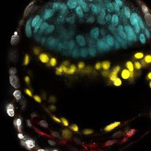
The First Human Embryo Model From Embryonic Stem Cells
Shelby Bradford, PhD | Mar 1, 2024 | 2 min read
Jacob Hanna developed a method for replicating embryogenesis outside of the uterus to understand the underlying mechanisms.
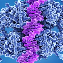
One Protein to Rule Them All
Shelby Bradford, PhD | Feb 28, 2024 | 10+ min read
p53 is possibly the most important protein for maintaining cellular function. Losing it is synonymous with cancer.
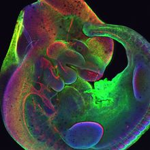
Illuminating Craniofacial Development
Hannah Thomasy, PhD | Jan 1, 2024 | 2 min read
Paul Trainor delves into the genetic and environmental factors that shape the head and face.
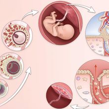
Infographic: Early Placenta Development Sets the Stage
Danielle Gerhard, PhD | Dec 4, 2023 | 2 min read
During early pregnancy, the placenta remodels the uterine environment to support fetal growth
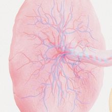
The Ephemeral Life of the Placenta
Danielle Gerhard, PhD | Dec 4, 2023 | 10+ min read
Recent advances in modeling the human placenta, the least understood organ, may inform placental disorders like preeclampsia.

Emerging from Silence: Capturing the First Heartbeat
Danielle Gerhard, PhD | Sep 27, 2023 | 5 min read
In the developing zebrafish, a noisy and asynchronous activity jumpstarts the heart’s journey to coordinated beating.
