The immune system is highly trained to detect and eliminate any potential threat to the human body. While years of evolution have turned this system into a pathogen-killing machine, the microbes it fights have also evolved intricate strategies to evade it.
A Parasite and the Art of Cloaking

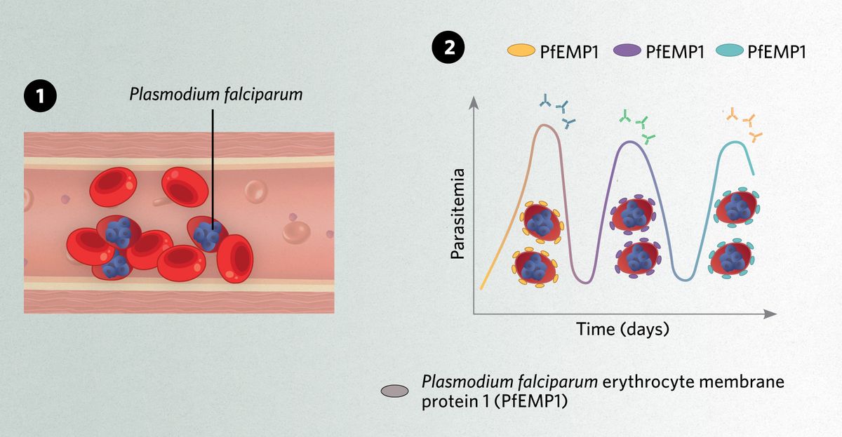
1) The malaria parasite Plasmodium falciparum expresses the protein PfEMP1 on the surface of erythrocytes to adhere the cells to blood vessel walls and escape clearance by the spleen.
2) PfEMP1 can be detected by immune cells. Through the process of antigenic variation, P. falciparum expresses different versions of it and escapes immune recognition.
Controlling the Enemy Within

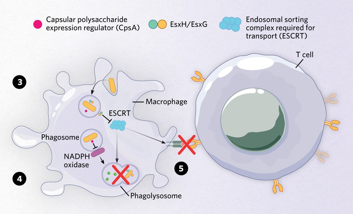
3) Inside macrophages, Mycobacterium tuberculosis dodges intracellular degradation by secreting virulence factors. Two effectors, EsxH and EsxG, inhibit the function of the ESCRT machinery, impairing the maturation of bacteria-carrying phagosomes.
4) Another Mycobacterium virulence factor, CpsA, disrupts another degradation pathway and blocks the activity of NADPH oxidase, impairing the destruction of the bacteria.
5) By affecting the normal function of ESCRT, M. tuberculosis EsxH-EsxG complex also disturbs the process of antigenic presentation via the MHC-II molecule.
A Viral Manipulator

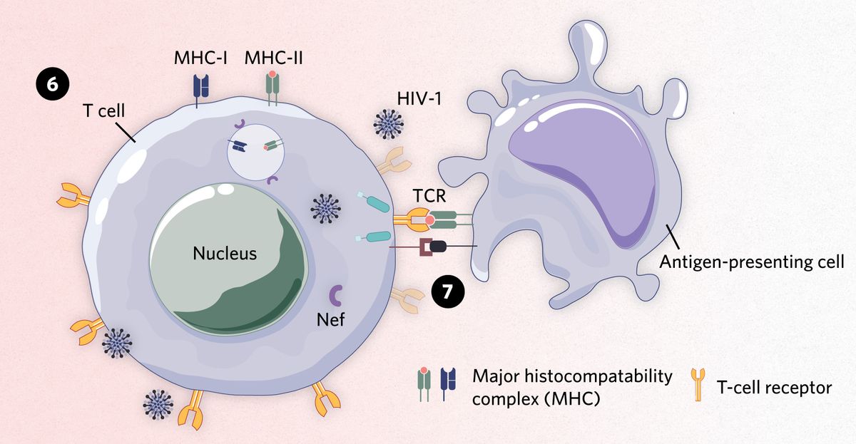
6) To stay hidden inside lymphocytes, HIV-1 expresses viral factors such as the negative factor (Nef) protein. In the infected cell, Nef downregulates the expression of MHC-I and MHC-II on the cell surface, impairing the presentation of viral antigens.
7) Nef also disrupts the proper formation of immunological synapses, the points of communication between T cells and antigen-presenting cells.
A Temporary Fungal Shield

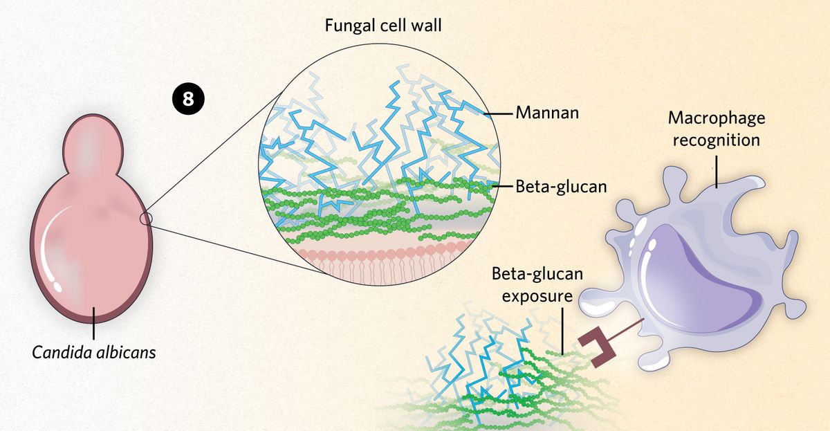
8) In Candida albicans, beta-glucans are a major target for immune detection by macrophages, which are one of the first lines of immune defense. The fungus covers its beta-glucans with a layer of mannans, shielding them from macrophage detection to prolong their stay in the host.
Read the full story.
- Deitsch KW, Dzikowski R. Variant gene expression and antigenic variation by malaria parasites. Annu Rev Microbiol. 2017;71:625-641.
- Chandra P, et al. Immune evasion and provocation by Mycobacterium tuberculosis. Nat Rev Microbiol. 2022;20(12):750-766.
- Fackler OT, et al. Modulation of the immunological synapse: A key to HIV-1 pathogenesis?. Nat Rev Immunol. 2007;7(4):310-317.
- Gow NAR, Lenardon MD. Architecture of the dynamic fungal cell wall. Nat Rev Microbiol. 2023;21(4):248-259.
- Gilbert AS, et al. Fungal pathogens: Survival and replication within macrophages. Cold Spring Harb Perspect Med. 2014;5(7):a019661.
