Live-cell imaging
In Living Color: Exploring Advances in Quantitative Live Cell Imaging
Leica Microsystems | May 28, 2024 | 1 min read
The latest technological developments and imaging platforms take live cell imaging to the next level.
Channeling Success: Simultaneous Multicolor Live Imaging
Leica Microsystems | May 21, 2024 | 1 min read
Researchers face many careful considerations when designing multiplex live cell experiments.
Illuminating Specimens Through Live Cell Imaging
Charlene Lancaster, PhD | Mar 14, 2024 | 8 min read
Live cell imaging is a powerful microscopy technique employed by scientists to monitor molecular processes and cellular behavior in real time.
Measuring Long-Term Neuronal Activity in Cell Culture
Sartorius | Dec 14, 2023 | 1 min read
Explore the latest advances in automated live-cell neuronal analysis for cell culture models.
Troubleshooting Fluorescence Microscopy Experiments
The Scientist and Evident | Nov 10, 2023 | 1 min read
Delve into the tactics used by scientists to overcome fluorescence microscopy’s greatest obstacles.
Striking a Balance for Perfect Images
The Scientist Staff | Oct 2, 2023 | 2 min read
Advanced microscope systems enable researchers to perform high-resolution live-cell imaging, while maintaining cellular health.
Observing Cells in Their Natural State with Digital Holographic Cytometry
The Scientist and Phase Holographic Imaging | Mar 1, 2023 | 3 min read
Technological and engineering advances let researchers delve deeper into cell function and behavior in physiological and pathological settings.
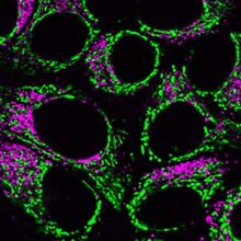
Choosing Fluorescent Reagents for Every Live Cell Application
The Scientist and MilliporeSigma | Nov 30, 2022 | 4 min read
Scientists gain unique insights into active biological processes with specific fluorescent probes, dyes, and biosensors.

Whole-Well Brightfield Cell Counting with the Latest in Microplate Imaging Technology
The Scientist | Oct 6, 2022 | 1 min read
In this webinar, Magdalena Eckschlager and Christopher Wolff will discuss how cutting-edge neural network-based algorithm technology enables label-free, live cell imaging and analysis.
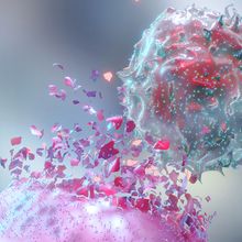
Phenotypic and Functional Characterization of CAR T Cells
Sartorius | Jan 19, 2022 | 1 min read
How advanced flow cytometry and live-cell analysis are accelerating immunotherapy research.

From Imaging to Kinetics: Measuring Calcium Signaling
Tecan | Sep 27, 2021 | 1 min read
Streamlining and automating cell-based assays for measuring intracellular calcium.
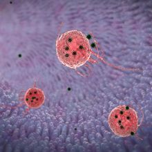
Technique Talk: Live-Cell Imaging Strategies to Quantify Phagocytosis
The Scientist Creative Services Team in collaboration with Sartorius | Sep 8, 2021 | 1 min read
Discover how to image and quantitate phagocytosis in real time
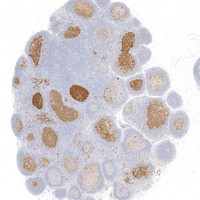
CAR T Cells Derived from Stem Cells Target HIV Tissue Reservoirs in Monkeys
Berly McCoy, PhD | May 25, 2021 | 3 min read
Transplanted CAR stem cells persisted long term and showed multilineage engraftment in tissues that harbor HIV.
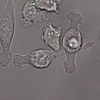
Engineered Immune Cells Eliminate Brain Cancer in Mice
Brooke Dulka, PhD | May 25, 2021 | 2 min read
Researchers developed a new CAR T-cell therapy that targets specific growth factor receptors in glioblastoma to eliminate brain tumors.
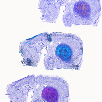
Environmental Cues Keep CAR T Cells on Track
Aparna Nathan, PhD | May 25, 2021 | 4 min read
Pairing CARs with a synthetic receptor makes T cells more lethal tumor killers.

Whole-Slide Imaging and Beyond: A New Open Platform for Scanning and Bioimaging
The Scientist | Oct 6, 2020 | 1 min read
In this webinar brought to you by MMI, learn how scientists use CellScan for a variety of lab applications, such as routine photo-documentation, whole-slide scanning, fluorescence imaging, time-resolved live-cell imaging, and combinations of these techniques.

Live-cell Imaging
The Scientist | Aug 27, 2020 | 1 min read
Download this eBook to take a closer look at past efforts, modern challenges, and future advances!

2019 Top 10 Innovations
The Scientist | Dec 1, 2019 | 10+ min read
From a mass photometer to improved breath biopsy probes, these new products are poised for scientific success.

Microscopy and Imaging Leader Shinya Inoué Dies
Jef Akst | Oct 7, 2019 | 1 min read
The long-time Marine Biological Laboratory scientist was known for using his own hand-built microscopes to image the dynamics of live cells.
