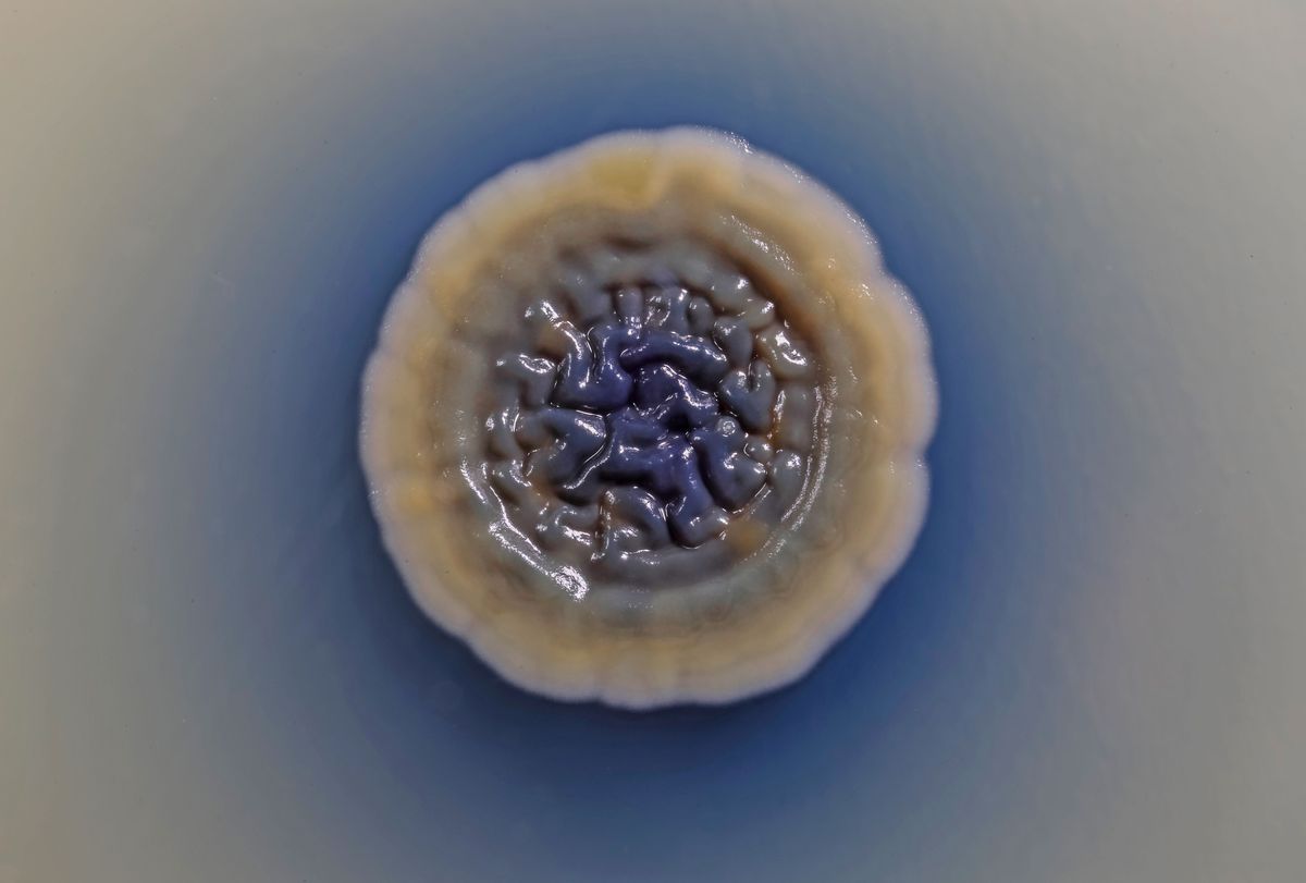
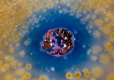
ABOVE: Microbiologist Scott Chimileski captures images of microbes such as Streptomyces coelicolor (center), which produces the blue antibiotic actinorhodin, inhibiting the growth of yellow Myxoccous xanthus colonies. Scott Chimileski, Marine Biological Laboratory
Microbiologist Scott Chimileski studies microbial biofilms at the Marine Biological Laboratory (MBL), exploring their intricate interactions and structural organization. His first brush with biofilms began during his graduate studies at the University of Connecticut, where he used confocal microscopy to capture time-lapse images of halophilic archaeal biofilms, which are relatively underexplored.1 “That’s where I really recognized the beauty of biofilms and their visual aspects,” he shared.
A lifelong interest in photography complemented his scientific pursuits, and the two passions merged fully during his postdoctoral work in Roberto Kolter’s lab at Harvard Medical School. Kolter, a fellow enthusiast for the artistic aspects of science, encouraged Chimileski to nurture this pursuit. “The two became totally, irreversibly intertwined,” Chimileski added.
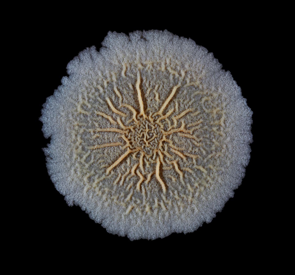
Ever since, Chimileski has captured the intricate structures of hundreds of bacterial biofilms at microscopic and macroscopic scales. Using a custom incubator camera setup, he created detailed time-lapse videos of colony growth, while electron microscopy allowed him to delve into cellular interactions at the micron level. This work resulted in a rich collection of detailed time-lapses. His extensive portfolio showcases bacteria with unique traits, using photography to share the beauty and complexity of microbes with scientists and the public alike.
Curating Microbial Exhibits in Museums
Chimileski’s passion for sharing science led to an interactive exhibition, Microbial Life: A Universe at the Edge of Sight, which he co-curated with Kolter. Hosted from 2018–2022 at the Harvard Museum of Natural History, the exhibit invited museum-goers—scientists and curious onlookers alike—to immerse themselves in the microbes found in everyday life and natural environments.

Visitors encountered familiar sights, such as a full-scale kitchen, and explored a live demo microscopy station with a rotating array of samples, from dust to kombucha. “I interacted a lot with the public,” said Chimileski. “The live demo station gave us a constant opportunity to introduce new topics and subjects. It was a constant source of new information.”
In another exhibit, Chimileski mounted four oversized Petri dishes onto the wall, each showcasing different living bacterial colonies. “Real growth and microbial evolution were happening inside these living Petri dish displays,” said Chimileski. “You could see mutants emerging at the edge of colonies and spreading faster.” For example, onlookers could watch a white Streptomyces colony slowly develop a rich halo of blue—an antibiotic called actinorhodin. “You’re seeing the natural pigment produced by the bacteria and the antibiotic production,” he explained. “This is important because half of our clinically-used antibiotics come from these soil bacteria.”
Booking a New Chapter: An Exploration of Microbes Around the World
Parallel to the exhibit, Chimileski and Kolter coauthored Life at the Edge of Sight: A Photographic Exploration of the Microbial World. This book highlighted a collection of Chimileski’s laboratory images and explored microbes’ roles in the history of the Earth and people’s lives. “One of the major themes of the book focuses on the macroscopic side of microbes,” he explained. “While individual microbes are microscopic, they manifest themselves macroscopically in all sorts of ways, from biofilms to these blooms of coccolithophores.”
Chimileski traveled to microbial science sites across the planet, describing these destinations as “Meccas of microbiology”. His journeys took him on hikes through the backcountry of Capitol Reef National Park to find stromatolites—fossils of cyanobacteria—and to the White Cliffs of Dover in the United Kingdom, where the iconic chalk cliffs are composed of algae bloom skeletons of coccolithophores.

Exploring Mouth Microbes Within Hedgehog Biofilms
After these projects concluded, Chimileski shifted his focus to studying biofilms in the oral microbiome, such as dental plaque, at MBL. “Biofilms aren’t just amorphous piles of bacteria, but the different species in these biofilms are localized relative to each other in recurring patterns or structures,” he explained. One such structure, called the hedgehog, features about 10 different bacterial species.2 These hedgehog communities, previously highlighted for their role in human health at the exhibit, offer a window into the intricate organization of microbial life.
At the heart of these hedgehog structures lies one bacterium called Corynebacterium matruchotii, which forms the central scaffolding.3 To study these structures, Chimileski cultured bacterial species together and used time-lapse imaging to observe their interactions. In one real-time experiment, he accidentally stumbled upon a unique way C. matruchotii divided. “It grew as a long filament at only one pole, called tip extension, which, in and of itself, is interesting. After it reaches a certain length, the cell divides simultaneously into 10 or more cells at once, which is called multiple fission,” remarked Chimileski. He contrasted this with binary fission, the textbook model for bacterial division, emphasizing that this unique behavior might have been overlooked using traditional fluorescence in situ hybridization methods, which require killing the cells.
Chimileski plans to continue capturing these images, delving deeper into the unseen behaviors of microbial communities. Looking ahead, he hopes to continue his science communication projects with exhibits and hopefully another book—possibly a 10th-anniversary edition—packed with more discoveries he’s yet to uncover.
- Chimileski S, et al. Biofilms formed by the archaeon Haloferax volcanii exhibit cellular differentiation and social motility, and facilitate horizontal gene transfer. BMC Biol. 2014;12:65.
- Mark Welch JL, et al. Biogeography of a human oral microbiome at the micron scale. Proc Natl Acad Sci U S A. 2016;113(6):E791-E800.
- Chimileski S, et al. Tip extension and simultaneous multiple fission in a filamentous bacterium. Proc Natl Acad Sci U S A. 2024;121(37):e2408654121.


